Home » Microscopy » RareCyte High Plex Spatial Biology System

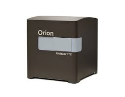
Orion is a novel technology and services platform offering the fastest path to
whole slide, high-plex imaging. Combining speed and subcellular resolution,
Orion enables comprehensive phenotypic profiling and characterization of
tissue architecture, tumor heterogeneity, and the immune response for whole
slide tissue sections in hours, not days.
Exceptional signal-to-noise ratio provides robust quantification of biomarkers
across a broad dynamic range. The ability to measure changes in expression of
low-level markers supports translational and clinical research.
Run panels from an extensive biomarker list, add in custom biomarkers, or
engage our services lab for custom panels and programs.

For comprehensive specifications, download the Orion specification sheet.
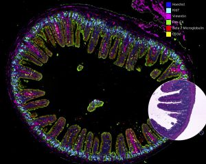
High-plex imaging across an entire slide, in a single scan, enables spatial analysis through examining biomarker expression, cell classification, and subcellular localization. Such analysis paves a path for advancing studies in pathology, immuno-oncology, infectious disease, and many more areas of disease.
Orion utilizes flexible hardware, software, and reagents to deliver the high performance, resolution and flexibility required for research and clinical assays.
By combining speed and resolution, Orion enables comprehensive phenotypic profiling and characterization of tissue architecture, tumor heterogeneity, and the immune response for whole sections in hours, not days.
Facilitate single round immunofluorescence staining and imaging of standard FFPE or fresh frozen tissue with the additional benefits of industry standard H&E and IHC modes. Run panels from an extensive biomarker list https://rarecyte.com/rareplex/, add in custom biomarkers, or engage our services lab for custom panels and programs
Orion technology breaks barriers, providing the fastest path to whole slide, highly multiplexed biomarker imaging data:
| High-plex, whole slide fluorescence imaging | Flexible panel design | ||
| Stain and image in hours, not days | FFPE and fresh frozen compatible | ||
| Rapid, single round stain and scan | Uses standard microscope slides | ||
| Subcellular resolution | Quantitative across a broad dynamic range | ||
| Same slide H&E imaging | OME-TIFF open file format |
Please fill out our form ,and we’ll get in touch shortly