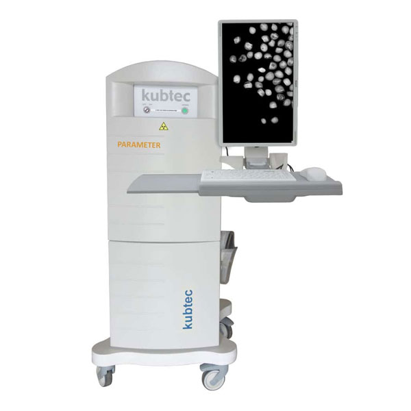
The KUBTEC PARAMETER® line is the company's cabinet X-ray platform for the research laboratory, running from straightforward high-resolution radiography up to multislice tomosynthesis. Across the line, Image Blender™ overlays the X-ray onto an optical photograph of the same specimen so the signal is read against real anatomy, and DIGIMUS Multi turns scans into quantitative bone mineral density and whole-body composition figures aligned with DXA methodology. Merkel Technologies supplies the complete PARAMETER range in Israel.
The KUBTEC PARAMETER® line is the company's cabinet X-ray platform for the research laboratory, running from straightforward high-resolution radiography up to multislice tomosynthesis. Across the line, Image Blender™ overlays the X-ray onto an optical photograph of the same specimen so the signal is read against real anatomy, and DIGIMUS Multi turns scans into quantitative bone mineral density and whole-body composition figures aligned with DXA methodology. Merkel Technologies supplies the complete PARAMETER range in Israel.
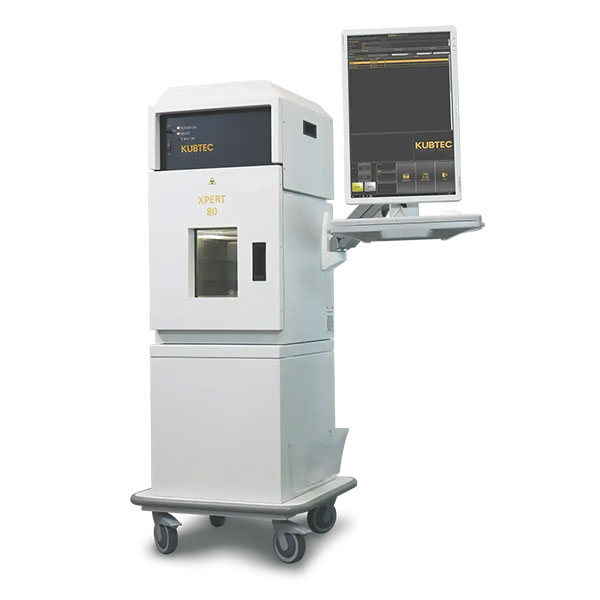
The KUBTEC XPERT® 80 line is the company's high-resolution cabinet X-ray platform, sitting alongside the PARAMETER systems and sharing their sub-50 µm resolution and DIGICOM specimen radiography software. Two models make up the line: the XPERT® 80 and the sharper XPERT® 80-L. Merkel Technologies supplies both in Israel.
The KUBTEC XPERT® 80 line is the company's high-resolution cabinet X-ray platform, sitting alongside the PARAMETER systems and sharing their sub-50 µm resolution and DIGICOM specimen radiography software. Two models make up the line: the XPERT® 80 and the sharper XPERT® 80-L. Merkel Technologies supplies both in Israel.
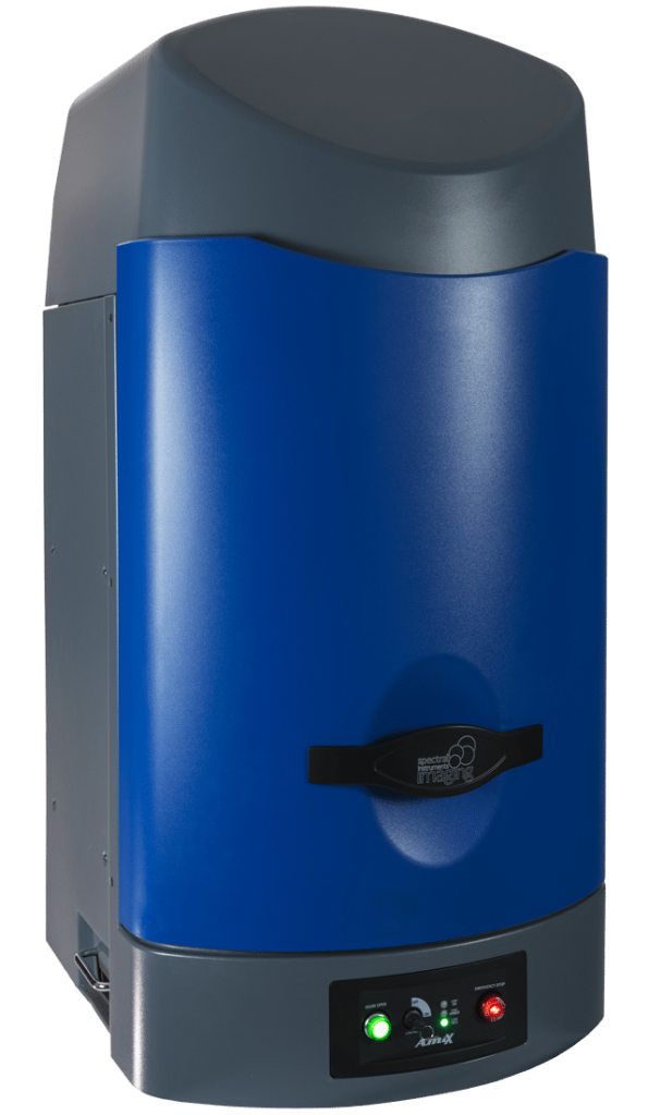
Fluorescence and bioluminescence is the most common method of pre-clinical in-vivo imaging. By using fluorescent or bioluminescent reporter genes it is possible to track gene expression, tissue growth, response to therapy, and much more applications.
Fluorescence and bioluminescence is the most common method of pre-clinical in-vivo imaging. By using fluorescent or bioluminescent reporter genes it is possible to track gene expression, tissue growth, response to therapy, and much more applications.
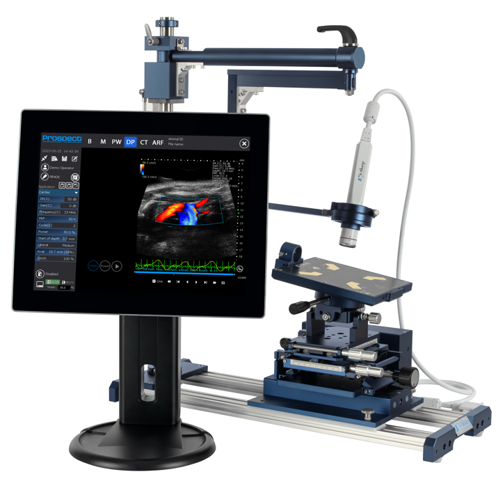
Ultrasound works by transmitting ultrasound waves and detecting the waves that bounce back from structures live images of soft tissue, vasculature, and organs are reconstructed. By using the doppler effect it is possible to measure to direction and speed of blood flow in blood vessels and the heart. Ultrasound is preferred for many applications due to its quick, real-time imaging with no required contrast agents or radiation. The pre-clinical ultrasound systems utilize high frequency to resolve small structures in rodents, including 6.5 days-old embryos.
Ultrasound works by transmitting ultrasound waves and detecting the waves that bounce back from structures live images of soft tissue, vasculature, and organs are reconstructed. By using the doppler effect it is possible to measure to direction and speed of blood flow in blood vessels and the heart. Ultrasound is preferred for many applications due to its quick, real-time imaging with no required contrast agents or radiation. The pre-clinical ultrasound systems utilize high frequency to resolve small structures in rodents, including 6.5 days-old embryos.