Home » Pre-clinical Imaging » Ultrasound Imaging
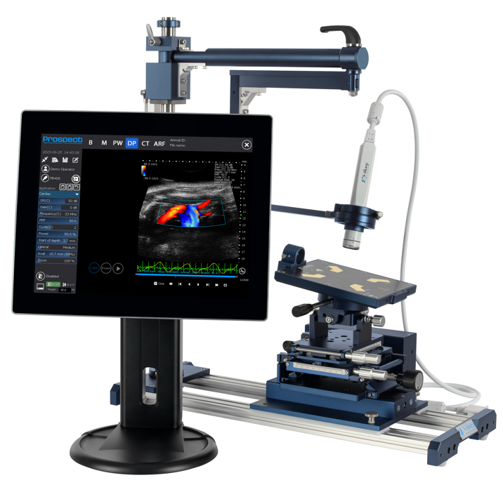
Ultrasound works by transmitting ultrasound waves and detecting the waves that bounce back from structures live images of soft tissue, vasculature, and organs are reconstructed. By using the doppler effect it is possible to measure to direction and speed of blood flow in blood vessels and the heart. Ultrasound is preferred for many applications due to its quick, real-time imaging with no required contrast agents or radiation. The pre-clinical ultrasound systems utilize high frequency to resolve small structures in rodents, including 6.5 days-old embryos.
Ultrasound is commonly used for organ size and blood flow measurement. For measuring the volume of tumors, the heart, kidneys, liver, and other organs the ultrasound will be used.
Blood flow measurements are commonly used for heart and kidney function as well as for tumor microenvironment.
Ultrasound is the only method for measuring blood flow in real-time and without the use of any contrast agents. The live imaging properties of Ultrasound allow measuring kinetics and biodistribution when using micro-bubbles as a contrast agent. Also, by using the speed of ultrasound propagation through tissue, it is possible to measure stiffness which can often indicate tissue fibrosis or other pathologies.
Please fill out our form ,and we’ll get in touch shortly