
Cell Imaging: Unveiling the Microscopic Universe Within
Introduction to Cell Imaging
The Evolution of Cell Imaging Technologies
Cell imaging techniques have evolved significantly throughout history. The journey of cell imaging began with basic optical microscopes in the 17th century, paving the way for the discovery of live cells by Robert Hooke. Advancements in staining techniques, notably with iodine and eosin in the late 19th century, allowed for improved visualization.
The 20th century witnessed the rise of fluorescence microscopy, utilizing fluorescent dyes and antibodies to target specific cellular components. Transmission electron microscopy, developed in the 1930s, enabled detailed imaging of internal cell structures at the nanoscale. Confocal microscopy, introduced in the 1950s, enhanced optical sectioning and three-dimensional imaging.
The 21st century brought revolutionary techniques such as super-resolution microscopy and phase contrast microscopy surpassing the diffraction limit and providing unprecedented clarity. Live-cell imaging techniques and sophisticated imaging modalities, like light-sheet microscopy and single-molecule imaging, further propelled cellular exploration. This historical progression highlights a continuous quest to unlock the mysteries of cellular biology through increasingly powerful and precise imaging tools.
Fluorescence Microscopy: Illuminating the Invisible
Principles of Fluorescence Microscopy
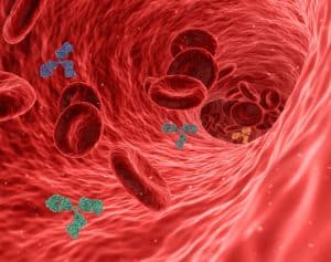
Fluorescence microscopy uses fluorophores, molecules that emit light upon excitation, to visualize specific cellular components. When exposed to a specific wavelength of transmitted light, these fluorophores absorb photons and re-emit them at a longer wavelength, producing fluorescent signals. This technique enables selective labeling and imaging of various cellular structures, proteins, or nucleic acids, providing insights into the dynamic cellular processes within living cells.
Applications in Biological Research
Fluorescence microscopy’s applications in biological research are extensive, encompassing studies of cell morphology, protein localization, and interactions and tracking cellular dynamics and behaviors. Additionally, it plays a crucial role in techniques like immunofluorescence, FRET (Fluorescence Resonance Energy Transfer), and live-cell imaging, contributing to advancements in fields such as cell biology, immunology, and neuroscience. The versatility and specificity of fluorescence microscopy have become indispensable for understanding complex biological phenomena at the microscopic level.
Confocal Microscopy: Capturing 3D Insights
Understanding Confocal Microscopy
Confocal microscopy uses a focused laser beam to illuminate a single plane of a specimen. At the same time, a pinhole aperture eliminates out-of-focus light, allowing for optical sectioning and enhanced resolution. This principle, known as confocality, produces sharper and clearer images than traditional microscopy. This process allows for more detailed imaging of a particular slice while minimizing contributions from out-of-focus regions. Confocal microscopy is crucial for obtaining clearer and more precise images.
Advancements in Imaging Resolution and Depth
Advancements in confocal microscopy have further improved imaging resolution. Techniques like spinning-disk confocal microscopy and multi-photon microscopy enhance speed and reduce phototoxicity, enabling live-cell imaging with improved temporal resolution. Super-resolution confocal techniques, such as stimulated emission depletion (STED) and structured illumination microscopy (SIM), surpass the diffraction limit, achieving resolutions approaching the nanoscale.
These innovations provide researchers with unprecedented details, facilitating the study of cellular structures and dynamics at higher precision. Confocal microscopy’s continual evolution has solidified its crucial role in biological research, offering remarkable imaging capabilities for a deeper understanding of cellular and subcellular processes.
Live-Cell Imaging: Witnessing Cellular Dynamics
Real-Time Observation of Cellular Processes
Live cellular imaging captures dynamic processes within living cells over time, offering invaluable insights into cell health, behaviors, responses, and interactions. This technique enables the observation of phenomena such as cell death, division, migration, and responses to stimuli in real-time. In drug discovery, live-cell imaging plays a pivotal role by providing a dynamic understanding of drug effects on cells, aiding in identifying and validating potential therapeutic compounds. It allows researchers to study drug pharmacokinetics, assess toxicity, and monitor cellular responses to candidate compounds.
Applications in Drug Discovery and Disease Research
In disease research, live-cell imaging contributes to understanding disease mechanisms, progression, and cellular responses to pathological conditions. This includes studying the behavior of cancerous cells, immune cell interactions, and neurodegenerative processes. The real-time visualization of cellular dynamics enhances our understanding of disease pathways and facilitates the development of targeted therapies. Thus, the Live cell imaging system is a powerful tool, bridging the gap between static snapshots and a dynamic understanding of cellular processes in drug discovery and disease research.
Advancements and Future Directions
Emerging Technologies in Cell Imaging
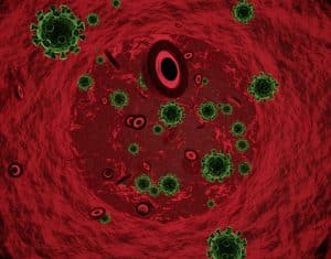
Super-resolution microscopy techniques surpass the diffraction limit of traditional light microscopes, providing unprecedented details at the nanoscale. Methods such as Stimulated Emission Depletion (STED), Structured Illumination Microscopy (SIM), and Single-Molecule Localization Microscopy (SMLM), including techniques like PALM and STORM, enable researchers to visualize cellular structures with exceptional resolution. These techniques break the diffraction barrier, allowing the imaging of structures as small as individual molecules.
Single-molecule imaging focuses on tracking and analyzing individual fluorescent molecules. This approach provides insights into molecular dynamics, interactions, and behaviors within living cells. By studying the movement and behavior of single molecules, researchers can unravel intricate cellular processes.
Applications of these techniques in cell imaging research are vast. They contribute to understanding cellular architecture, protein localization, and interactions, aiding research in areas like neurobiology, immunology, and cell signaling. Super-resolution microscopy and single-molecule imaging have revolutionized cell imaging by offering unparalleled insights into the nanoscale world of cells.
FAQs About Cell Imaging
What is the difference between fluorescence microscopy and confocal microscopy?
Fluorescence microscopy and confocal microscopy are related techniques, both utilizing fluorescence to visualize cellular structures, but they differ in their imaging capabilities and principles. Fluorescence microscopy relies on the ability of certain molecules, called fluorophores, to emit light of a specific color when exposed to a particular wavelength. Confocal microscopy also utilizes fluorescence but incorporates a pinhole aperture to eliminate out-of-focus light, resulting in improved resolution and optical sectioning.
How does live-cell imaging differ from traditional cell imaging techniques?
Live-cell imaging is specialized for observing dynamic cellular events in real time, while traditional cell imaging techniques focus on static snapshots for studying cellular structures and morphology. Each approach has its unique advantages and applications in cellular research.
What are some challenges associated with live cell images?
Live-cell imaging poses several challenges due to the dynamic nature of live cells and the need to maintain cell viability during the imaging process. Some of the key challenges include:
- Phototoxicity
- Cell Viability
- Autofluorescence
- Bleaching
- Movement and Drift
- Limited Observation Time
- Data Analysis
Researchers continuously work to address these challenges through advancements in microscopy technologies, imaging protocols, and the development of new fluorescent probes and imaging methodologies to enhance the reliability and biological relevance of live-cell imaging.