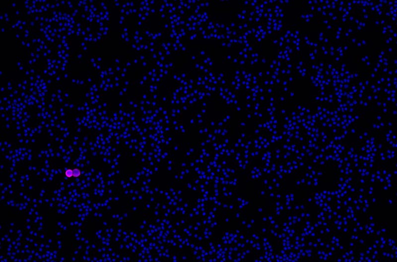
RareCyte enables practical deployment of circulating tumor cell (CTC) and rare cell-based liquid biopsy with instrumentation and consumables that provide an exquisitely sensitive, accurate, reproducible, and transparent workflow from blood collection to single cell isolation.
https://link.springer.com/article/10.1007/s10549-022-06585-5

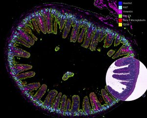
Orion is a novel technology and services platform offering the fastest path to
whole slide, high-plex imaging. Combining speed and subcellular resolution,
Orion enables comprehensive phenotypic profiling and characterization of
tissue architecture, tumor heterogeneity, and the immune response for whole
slide tissue sections in hours, not days.
Exceptional signal-to-noise ratio provides robust quantification of biomarkers
across a broad dynamic range. The ability to measure changes in expression of
low-level markers supports translational and clinical research.
Run panels from an extensive biomarker list, add in custom biomarkers, or
engage our services lab for custom panels and programs.

For comprehensive specifications, download the Orion specification sheet.
Orion is a novel technology and services platform offering the fastest path to
whole slide, high-plex imaging. Combining speed and subcellular resolution,
Orion enables comprehensive phenotypic profiling and characterization of
tissue architecture, tumor heterogeneity, and the immune response for whole
slide tissue sections in hours, not days.
Exceptional signal-to-noise ratio provides robust quantification of biomarkers
across a broad dynamic range. The ability to measure changes in expression of
low-level markers supports translational and clinical research.
Run panels from an extensive biomarker list, add in custom biomarkers, or
engage our services lab for custom panels and programs.

For comprehensive specifications, download the Orion specification sheet.
Nanolive’s imaging and analysis platform – the CX-A – is the only solution in the world to offer automated 3D label-free live cell imaging that is compatible with 96-well plates.
* Non-invasive autofocus solution: keeps cells in focus for the time of the experiment.
* Integrated image analysis solution with multi-parametric read-out.
* Long-term imaging of living cells with no perturbations and no contamination in physiological conditions thanks to our dedicated incubation solution. More details here.
* High quality motorized stage with a micron range precision repeatability of motion.
* Stitching feature: allows to analyze highly confluent cell populations containing hundreds of cells while keeping Nanolive’s signature sub-cellular resolution.
* Meaningful correlative imaging: features a fully integrated 3 channel epifluorescence imaging modality.